
ultrastructure: a parable in human understanding
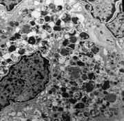
Recently I rotated in pediatric pathology. One day the attending was nice enough to go over images of tumors obtained under the electron microscope with us. This technique allows visualization of sub-cellular structures such as organelles that may have diagnostic impact on the classification of tumors.
It is interesting to note that electron microscopy ("ultrastructure") is rarely used in diagnostic pathology anymore. However, in the past it was considered so important that volumes were published, even in subspecialities such as "electron microscopy of the gastrointestinal tract". Looking through one such book, I saw entire chapters devoted to morphological variations of mitochondria, and was reminded of the book "On Growth and Form", a mathematical study of biological forms published in the newly-post-Darwinian year of 1917 (with many pictures detailing the geometry of nature -- and this was before fractal geometry was discovered).
In retrospect, the medical phenomenon of electron microscopy arose from a solid belief in reductionism. Thas it, the light microscope only afforded a limited capacity to zoom in on biological structures. If the most basic cellular components could be visualized (with electrons), then it was felt one could know from what tissue the tumor cell arose. Armed with this knowledge cancer could be most appropriated treated, even those forms that seemed ambiguous under light microscopy.
The problem is that there turns out to be few instances in which electron microscopy actually yields useful diagnostic information. Like the current studies into DNA analysis, it remained an expensive speciality that over the course of years failed to provide the masses of useful information expected. So pathology returned to the mainstay pioneered centuries ago: thin slices of human tissue stained on glass slides, examined under a light microscope. The study of human tissue in this fashion is known (rather derisively today) as "morphology".
The latest diagnostic technique in vogue is immunohistochemistry, where glass slides of human tissue are specially stained with colored compounds attached to antibodies, so that specific cellular receptors indicative of cell type can be visualized. This has become so widespread that many new pathologists do not properly learn the basics of morphology, and instead focus on the immunohistochemical profile of various tumors.
It would be great if immunohistochemistry ("immuno") worked perfectly, but unfortunately many of the stains are not as specific as originally thought. This basically means that many cells will be positive for a stain originally thought to only highlight one type of cell. Even when numerous stains are used together in a panel, many tumors still refuse to fit into the box, and this represents an area of great interest in pathology.
So what happens to the tumors that still can't be classified with immuno? We go back to the old-school diagnosis, based on medical history, site affected, and histological appearance, and try our best.
It is interesting to note that tumors are classified in a taxonomical system similar to the one used by Linnaeus to classify life forms. The basis for understanding rests on what type of tissue is deprecated and begins reproducing in an uncontrolled fashion. Therefore there are tumors of muscle, tumors of skin, tumors of bone, etc. This sounds good, but some tumors have cells that don't really resemble any particular normal tissue. Worse yet, some tumors transform and instead resemble a different tissue than the one they came from. It begs the question, how accurate is it to think in terms of taxonomy?
In fact, any classification scheme is really an intellectual artifact wrapped up in suspension of disbelief. As humans, we like to categorize things to convince ourselves that we understand them. When something does not fit into any particular category, we create a new category for the thing and try to find aspects that characterize it. The entire system is based on differences and similarities, and yet no one really knows the criteria for how different two things must be in order to be considered from a different category. What if they are just variations on a common ancestor?
In Wikipedia, under the entry "Taxonomy" there is a link to "Numerical taxonomy" which is also called "Data clustering". This is the study of partitioning things into groups based on statistics; a field most pathologists have probably never heard of. Maybe this is the future of a new way to think that better classifies what we see and how to use that classification system. But in the meantime I suspect most young pathologists are so caught up in tenure track and DNA-chips that they don't pause to consider the underlying premise they operate upon.
David MacCauly wrote and illustrated a book about archaelogists who in the future excavate the remains of a typical American city. But the future archaelogits base many of their assumptions on faulty conceptions, and end up surmising that billboards along the highway are totems to religious deities and that the bathroom is a place of worship. It makes me wonder about a future time when my colleagues will have entire sections of their bookshelves devoted to tomes on immuno and molecular biology, and some pesky resident will wonder about the strange acronyms and colorful diagrams meant to represent... just exactly what?
posted by John Kacher | 9:58 AM
![]()
0 Comments:
Post a Comment
<< Home